


Spinal Vascular Malformation Embolization
What is a Spinal Vascular Malformation (SVM)?
A spinal vascular malformation (SVM) is a congenital abnormality of spinal blood vessel development resulting in a direct communication between arteries and veins. There are a wide variety of types of SVMs ranging from an abnormal communication along a nerve root, within the spinal cord itself, along the surface of the spinal cord, or diffusely involving a given spinal segment. Different types of SVMs have both different management approaches and prognosis depending on the specific factors in any given case.
What are the symptoms of an SVM?
SVMs may present in a variety of ways depending on the type, ranging from myelopathy (weakness and numbness) to pain. Symptoms may develop from shunting of blood in which portions of the spinal cord may not receive adequate blood supply or if the venous side is pressurized and cannot drain normally. Bleeding from the malformation into the spinal cord itself or the surrounding fluid space may also occur.
How is an SVM diagnosed?
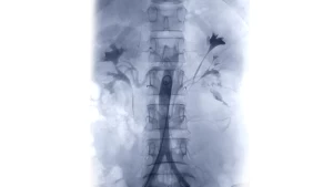
Spinal vascular malformations are typically diagnosed on an MRI scan, then further defined by a catheter angiogram.
How is an SVM treated?
As mentioned above, SVMs, while rare lesions are very heterogeneous in terms of their location, architecture, blood supply, and symptoms. The two main treatment modalities are endovascular or surgical. For an endovascular procedure, very careful spinal angiography is necessary to define what vessels are supplying the malformation and what vessels are supplying to the normal spinal cord. If this is adequately delineated, embolization may be performed with glue (NBCA) or a glue-like material (Onyx) to block off the abnormal connection. If this cannot be performed safely from a catheter approach due to the blood supply, open surgery may be necessary to place a clip on the abnormal communication without placing other blood vessels or the spinal cord at risk. Each case demands very specific tailoring of a multidisciplinary treatment plan.